
Fungus
-
Posts
2,833 -
Joined
-
Last visited
Content Type
Profiles
Forums
Classifieds
Tip Site Directory
Blogs
Articles
News
Arborist Reviews
Arbtalk Knot Guide
Gallery
Store
Freelancers directory
Everything posted by Fungus
-
Which means they are associated with Carpinus and not with Corylus.
-
Guy, 1. I'm not that old, but I've known the tree for 45 years, as us boys in our teens in the early days of courtship took our girlfriends to a still existing little stone "gazebo" in the woods close to this impressive beech. The tree was split in half (torsion forces) and fell two years ago in a storm, which uprooted 100+ oaks standing beside local roads within the region, where I have returned to, after 8 years of living and working in the German Eifel, three years ago. I've re-assessed and monitored this forest since my return and compare the data with the data I collected 28-25 years ago, when we spent some time in a summer house nearby. 2. The splitting in half was done by the mycelial sheets of the simultaneous white rotter Fomes fomentarius, which had colonized the trunk of the tree from 3 metres up from the forest floor and for the greater part was (partially panic) fruiting after parts of the trunk and the crown branches where horizontal with soil contact. The wood decay (white rot with selective delignification) at and just above ground level was coming from the mycelium of G. lipsiense (= G. applanatum), which made the tree especially vulnerable for wind throw, which probably would have happened if the tree wasn't split in half. Also see Tony's (Hamadryad) post. 3. No, until recently, in The Netherlands there was a saying : with thunder and lightning about, avoid (acid rich) oaks, which attract lightning and find shelter underneath the canopy of a beech, because beeches were never struck by lightning. Nowadays beeches standing at the edges of woodlands, which are heavily under the influence of ammonia deposition (nitrification) coming from maize fields and manured grasslands, are sometimes struck by lightning because of the changes in the constituents of the surface of their "skin" (bark), which turn the bark from neutral into negative attracting electrical discharges of lightning because of the hightened conductivity. 4. I suppose, you mean Francis Schwartze, a former employee of the "VTA-professor" Claus Mattheck, I did field research for and worked with for 10+ years. Schwartz's research into Fomes and Ganoderma species was for the greater part done under controlled in vitro or laboratory settings. The checking of his findings in field studies with life trees was done by German and Dutch tree health specialists and a mycologist (me). If you want to read more about the differences between the tree species specific attacking strategies of the perennial necrotrophic parasitic G. lipsiense and the perennial biotrophic parasitic G. australe (= G. adspersum), I suggest you digest what can be found on this forum if you use Search and scan for the Ganoderma thread and other posts and threads with information on (other) Ganoderma species, such as the annual G. lucidum, G. resinaceum or G. carnosum and the perennial G. pfeifferi. 5. No violent past (see 1.), but entirely attributable to fungal activity - especially of Fomes - inside the tree.
-
Yes, but primairely under oak. And on old blackened FB's, one can often find Nyctalis asterophora (=Asterophora lycoperdoides) and sometimes N. parasitica.
-
David, Nice pictures ! Two remarks, the FB of R. fragilis looks far from fragile to me, so did you look for the characteristic serrate or "saw toothed" edge of the gills ? Did the small greenish FB to the left belong to the same species/mycelium ? If so and without the serrate margin of the gills, but with buttery and not crumbly gills instead, could it be R. cyanoxantha s.l. ? And it's R. atropurpurea.
-
Cassian, Photo 1 : without pores and tubes I would expect this to be a corticiod Aphyllophorales, with pores and tubes a poroid Aphyllophorales. Photo 2/3 : without microscopical analysis this looks like mycelial/hyphal sheets of a not yet fruiting macrofungus. Photo 4 : Anamorph of what seems to be K. deusta. Photo 5 : Teleomorph of the same species. I didn't find the photo of the Orange FB's you promised, which presumebly would be of the Cryphonectria. As far as conclusions can be reached in this phase of the analyses and with the present macroscopical documentation, I would attribute the dramatic effects to a combined strategy of the Cryptonectria and K. deusta, where the Cryphonectria is the primary parasitic and not wood degrading pathogen and K. deusta is the secondary pathogen starting off as a saprotrophic soft rotting the heart wood inside out and becoming parasitic once the living tissue is reached through invasion of radial rays and the remaining living cambium is killed, after which it fruits with anamorphs within a year developing into teleomorphs. If this is the case, the FB's of Cryphonectria should be found scattered over the trunk (causing local bark and cambium necrosis and/or canker ?) and the FB's of K. deusta should be found at the base of the trees.
-
Guy, In this case, Chondrostereum purpureaum is not a pathogen, but a (secundary) saprotrophic and the infection took place via everywhere and ever present airborn spores.
-
Guy, 1. That's were we (again) disagree. There was a lack of scientific validity in the methods you used, as the data were gathered from a very limited and not adequately assessed and documented field "research" perspective, which may have been adequate for the tree owner, but does not qualify for being presented as a scientific contribution to the subject. 2. See : Forum Training & Education : Mycological Tree Assessment. 3. I didn't retract, I wrote : "Phytophthora (in general), which includes P. ramorum. 4. Just like a few other senior members of this forum, I have a "mission" to fullfill, so no teaching without preaching. 5. Let me remind you, that it was you, who "invaded" my thread challenging my research on the subject and uploaded an article from 2004, which made me assume, that you wanted your article and claims reviewed from my mycological and forest ecological perspective and by my scientific standards. And concerning your next post on the "baby tree". By what method did you asess the constituents of the "frothy flux" and exclude Phytophtora ramorum (SOD) or Acute Oak Death being responsible for the ooze ?
-
David, - R & G only give size ranges for G. carnosum : 1-10 x 0.7-4 cm, so do B & K : G. carnosum 5-25 x 1-4 cm. - Ellis & Ellis give size ranges for G. lucidum : up to 25 x 3 cm and for G. carnosum : up to 25 x 4 cm, - Jülich for G. lucidum : 5-15-25 x 1-2.5 cm and for G. carnosum : 1-10.5 x 0.7-4 cm, - Jahn for G. lucidum : up to 25 cm, - Däncke for G. carnosum (and implicit for G. lucidum) : up to 8 x 2 cm, - Phillips for G. lucidum : up to 24 x 1-3 cm, - and Donk for G. lucidum : up to 20 cm.
-
Any details on the fungus in photo 4 and 5 : gills and spore colour, stipe central or excentric ?
-
David, on the sporee, you and Tony were right then , so even though with non-species specific annual FB's, convincingly speeking for Ganoderma ! But what is your opinion on both species being (long) stalked, as stated by both Breitenbach & Kränzlin and Ryvarden & Gilbertson, or should we regard and list these specimen as typical for G. carnosum on yew, its main host in the U.K. ?
-
Which still needs to be proven on a piece of white or black paper and not on a (beech) leave or piece of wood, which is brown by itself.
-
Guy, My apologies too . No offence meant, just correcting invalid methods of assessment or (field) "research" and jumping to conclusions based on non-scientific data derived from a limited perspective, that's all, so also an even shorter reply in return. 1. Wrong conclusion. As a forest ecologist and mycologist, my focus is on the total tree species specific ecosystem and its soil food web, including relevant parasitic, saprotrophic and symbiotic macrofungi and other organisms, not on the trees, nor on the fungi alone, but on all of the contextual or "holistic" (Gestalt) (eco)system aspects (habitat, niche), without which IMO a complete and valid analysis of the health or condition of a tree can not be made. 2. You can see and identify 1-2 µm wide hyphae with a hand lense ? 3. Correct, you said Phytophthora (in general), which includes P. ramorum. 4. I thought, that quotation marks or inverted comma's are not only used for citations, but that a word between "..." in both languages also means : not literally to be understood as such. So let's agree to disagree and leave it at that .
-
Funny enough are all the reports on G. carnosum elswhere in Europe mostly on Abies and sometimes on Taxus, Larix, Picea, Pinus or Pseudotsuga and also on Betula, Carpinus, Fagus or Quercus and is G. lucidum also reported from Picea, which is the host in half of the cases found in Fennoscandia. So one starts wondering how many of the findings of (stalked) G. carnosum and G. lucidum are identified correctly.
-
Also from a visual perspective, the - non-stalked - FB's in the photo's look far to irregular or "wülstig" to be an annual Ganoderma, whether it is G. carnosum, G. lucidum or G. resinaceum and the pores look much more like the (irregular) pores of H. annosum, which are just as big or even a bit smaller then those of G. carnosum. So the only decisive characteristic left is the colour and - provided it is a Ganoderma species - the size of the spores.
-
Tony, Did you ever see G. carnosum "in the flesh" and what literature do you use ? Breitenbach & Kränzlin : G. carnosum pores 3-4 per mm, H. annosum pores 3-4 (5) per mm ; Ryvarden & Gilbertson : G. carnosum pores 3-4 per mm, H. annosum pores 4-5 per mm, so how much larger are the pores of H. annosum then ? You even have started me wondering whether the identification of Guy Watson's Ganoderma on yew was correct, as all the photo's of G. carnosum in books and on the internet show annual FB's with well developed, according to Ryvarden & Gilbertson and Breitenbach & Kränzlin, 5 to 10-25 cm long stipes, looking like an upright threatening cobra, just as G. lucidum mostly does.
-
Tree assessment/forest pathology
Fungus replied to Tony Croft aka hamadryad's topic in Fungi Pictures
Tony, If the criterium for being a winter fungus would be having pictures of a species with snow on top, then the Fly Agaric would also be a winter species, as I found it once on the 20th of December with white velum warts and lots of frozen snowflakes on top. And in my calendar, late fall preceeds winter time. The decisive characteristic is having trehalose, a kind of anti-freeze, in the cells of its FB's, protecting the cells and especially the reproductive organs from freezing (exploding) and making the revival of the FB's possible, so that reproduction can restart after the frost is gone, which only is the case for two species, i.e. P. ostreatus and F. velutipes. -
... and has a distinctive unpleasant sour sweet smell, which also cannot be used for ruling out a species from a picture .
-
Tree assessment/forest pathology
Fungus replied to Tony Croft aka hamadryad's topic in Fungi Pictures
That's the kind of misunderstanding one gets when not using the Latin names, as in Dutch, P. serotinus is called Groene schelpzwam, which means Green shell (i.e. not oyster) fungus. So then there is no connection to Armillaria, as I before described for P.ostreatus, because P. serotinus can not survive longer lasting frost periods and thus is not a winter species such as P. ostreatus and Flammulina velutipes are. -
Tony, I've seen lots of Heteroporus annosus brackets looking exactly like these on several coniferous tree species, including yew, and even on beech and Euonymus europeus, so only the colour of the spores will be decisive in this case.
-
Tree assessment/forest pathology
Fungus replied to Tony Croft aka hamadryad's topic in Fungi Pictures
Tony, 1. No, I've never seen the Nectria/Cryptococcus combination "rising up" from and fruiting at the base of the trunk. IME this type of body language on beech is always caused by Armillaria coming from an infection of the roots and/or buttresses. 2. Correct, with the addition, that the mycelium of P. ostreatus needs a frostbite to start fruiting and "uses" the already poorer condition of the tree and the tree's resting period during wintertime to attack the living tissue and decompose dead wood without the tree being able to defend itself. -
Rob, Well noticed . Annual sterile panic fruiting of the nearly dead mycelium of G. lipsiense at this side of the base of the trunk.
-
Tree assessment/forest pathology
Fungus replied to Tony Croft aka hamadryad's topic in Fungi Pictures
An example of the body language of the next phase of an infection with a parasitic Armillaria species in beech. ---
-
And for those interested in the Mycena in the fifth photo with the old Gano's, it's M. galericulata, a species decomposing wood with organohalogenes or polyaromatic hydrocarbons.
-
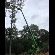
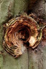
Documentation of a beech, which has been split in half by the mycelia of Fomes fomentarius and Ganoderma lipsiense, is provided. The about 150 years old beech is standing in a mixed forest, which has not been managed over some decades, on poor sandy soil. Photo 1/2/3 : show the remaining parts of the trunk from two sides after the tree has been split in half and lost its crown, with the branches falling towards two sides of the tree trunk. Photo 4/5 : show the inactive and active perennial brackets of G. lipsiense on two sides of the base of the trunk. Photo 6/7 : show the perennial brackets of F. fomentarius on the broken off upper trunk, which have for the greater part been developed after the tree was completely delaminated, split in half and the crown fell to both sides of the remaining trunk. Photo 8 : shows the "brittle" white rot with selective delignification bordered by black demarcation lines caused by the mycelium of G. lipsiense in a piece of central wood from the base of the trunk.


-
Documentation on an about 70 years old beech with extreme sun scald and loss of bark is presented. The beech is standing beside a busy road. Until spring last year it was protected against sunlight overexposure by two Tilia's, which were pollarded heavily March 2010, standing opposite to the south side of the beech. This year about half of the bark fell off after the dead cambium was decomposed by the mycelium of Chondrostereum purpureum, which FB's are all over the bark of the tree trunk. Photo 1 : shows the beech opposite to both pollarded Tilia's. Photo 2 : shows the south side of the beech facing the Tilia's. Photo 3/4/5/6 : show the patches of bark coming off the trunk. Photo 7/8/9 : show the patches of bark falling from the major branches of the crown. Photo 10 : shows the FB's of Chondrostereum purpureum. ---